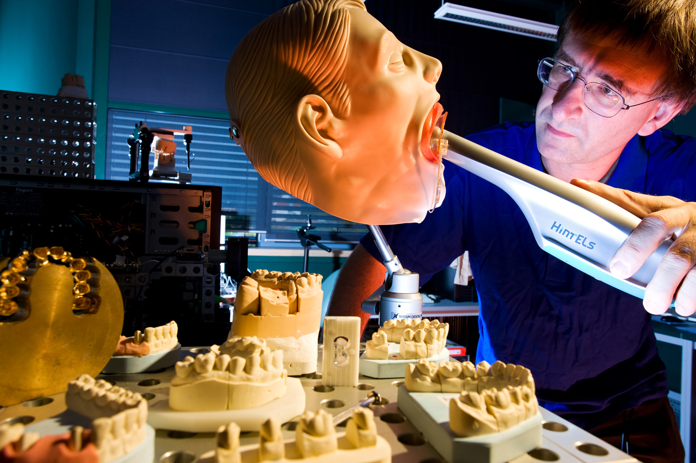
Dental snapshot
At present, dental technicians can only make dentures using a bite impression. The silicone template for this plaster model is made by the dentist, in a procedure which is unpleasant for the patient. In future a 3-D digitizer will provide the teeth contours – without plaster model.
When toothache makes a visit to the dentist unavoidable this often marks the start of a time-consuming treatment marathon for the patient. If the tooth cannot be saved and a dental prosthesis is necessary, the dentist first has to make a silicone impression for the dental laboratory. The patient is sent home with a provisional repair and dental technicians set to work on modeling a plaster impression. The model is then scanned using digital cameras and from the geometric measurement data obtained the matching dental prosthesis is produced.
The intricate and laborious route from bite impression and plaster mold to model scanning in the laboratory could soon be a thing of the past. “The three-dimensional coordinates of the tooth surface can be determined on the basis of measurements taken in the patient’s mouth,” says Dr. Peter Kühmstedt, group manager for 3-D measurement technology at the Fraunhofer Institute for Applied Optics and Precision Engineering IOF in Jena.
Under a contract from German dental company Hint-Els, an expert team at the Fraunhofer institute developed an optical digitization system which scans the oral cavity and captures three-dimensional data of the teeth using camera optics. A complete picture of the individual tooth is created from several data records. After an all-round measurement, it is even possible to represent the complete jaw arch as a virtual computer image. The measurement conditions in the confined oral cavity are, however, unfavorable. To obtain precise results, the scientists use fringe projections in which a projector shines strips of light on the tooth area to be measured. From the phase-shifted images the evaluation software determines the geometric contour data of the tooth. Two camera optics provide the sensor chip with image information from different measurement perspectives. After the pixel-precise comparison of various camera images, the evaluation program recognizes any image faults and removes them from the complete image.
It is problematic if the patient moves while the images are being taken in the oral cavity. The scientists have therefore made sure that the process takes place quickly. “The image sequence for each measurement position is captured in less than 200 milliseconds,” explains Kühmstedt.
