Fraunhofer-Gesellschaft
Making the invisible visible with entangled quanta
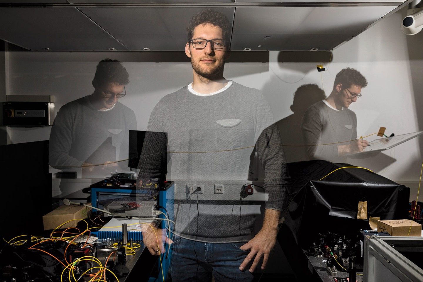
Dr. Markus Gräfe and Marta Gilaberte Basset, are pressing entangled photons into service for quantum imaging, a field that scientists just recently began to explore. “Medical researchers have been stymied by the problem of human cell samples being so sensitive to light. UV light, especially, damages cells. This limits the amount of time available for investigation,” says Gilaberte Basset. Quantum imaging with minimal doses of radiation could allow scientists to observe and analyze tissue cells with a high resolution and non-destructively. They could even monitor cell processes that take several minutes or many hours.
Blue laser beams flash this way and that among the Jena lab’s elaborate apparatuses as Gräfe explains the gist of their research: “We use entangled photons covering the entire optical spectrum from infrared to ultraviolet in our microscopic systems. This enables us to make objects visible even at wavelengths that had been invisible to date. To put it simply, the light beam that probes the object is not the same light used by the camera to take pictures. While one set of photons is sent to the object to be detected at invisible wavelengths, the twin photons in the visible spectrum are captured by a camera. The entangled light particles carry the same information, so this generates an image despite the fact that the light reaching the camera has never ‘seen’ the actual object.”
Quantum effects of this nature fascinate physicist and space researcher Marta Gilaberte Basset. “The quantum world is difficult for our imagination to grasp. Nonetheless, we experience its laws in action every day in the lab. At the end of the day, this is all about allowing for the existence of two different entities.” Basset believes in the viability of practical applications, for example, high-quality imaging to help diagnose cancer. She is exploring that possibility in her doctoral thesis.
The IOF research team documented its enthusiasm for applied quantum mechanics with the world’s first quantum video. Their choice of subject was inspired: They filmed a smiley without actually pointing a camera at it. It seems this emoticon was an accurate reflection of the researchers’ state of mind when they achieved this milestone in quantum imaging.
Real-world applications are a top priority for the IOF team. Markus Gräfe says that integrating quantum technology into today’s microscope systems is a key challenge. He wants to do this in a way that brings down the barriers for industry: “Nobody wants to fuss with several systems or work with one oversized system. This is why we’re keeping the communication with microscope makers such as Carl Zeiss flowing and are working together on practicable solutions.” The IOF is also pooling its research into quantum imaging with the expertise of five other Fraunhofer Institutes. This lighthouse project goes by the name of QUILT – Quantum Methods for Advanced Imaging Solutions. Several companies have joined the consortium’s industry advisory board in a bid to keep its efforts to develop quantum-based imaging, spectroscopy and analytics solutions on an application-oriented track.
Gräfe, Gilaberte Basset and their Fraunhofer colleagues have already achieved tangible results. One is a portable demonstrator microscope for life-science applications. Another is the first quantum spectrometer for the midrange infrared band developed by a research team at Fraunhofer IPM in Freiburg that is also taking part in QUILT. Medical science is not the only field that stands to benefit from quantum technologies. Similar approaches could work in fields such as materials science. Automakers, for example, would welcome better detectors that inspect paint layers in the terahertz range in far greater detail.