Quantum imaging

Quantum imaging: using quantum tricks to identify tumor tissue

Goebel’s colleague at Fraunhofer IOF, Dr. Karin Burger, is also working on entangled light particles with her project team. Instead of working on improving secu­rity against eavesdropping, though, she is focusing on medical diagnostics. During an operation, the surgeon often does not know whether the entire tumor has in fact been removed. A sample taken from the tissue at the margin is sent to the lab, where lengthy contrast methods are combined with optical microscopy to tell diseased and healthy cells apart. This takes time and can result in a need for follow-up procedures. “Newer digital microscopes with infrared detectors don’t need additional fluorescent markers, but their limited signal-to-noise ratio means they can quickly reach their limits. And higher-resolution systems are very large and require additional cooling,” Burger explains. This is why medical centers such as Jena University Hospital are looking for faster, more efficient methods.
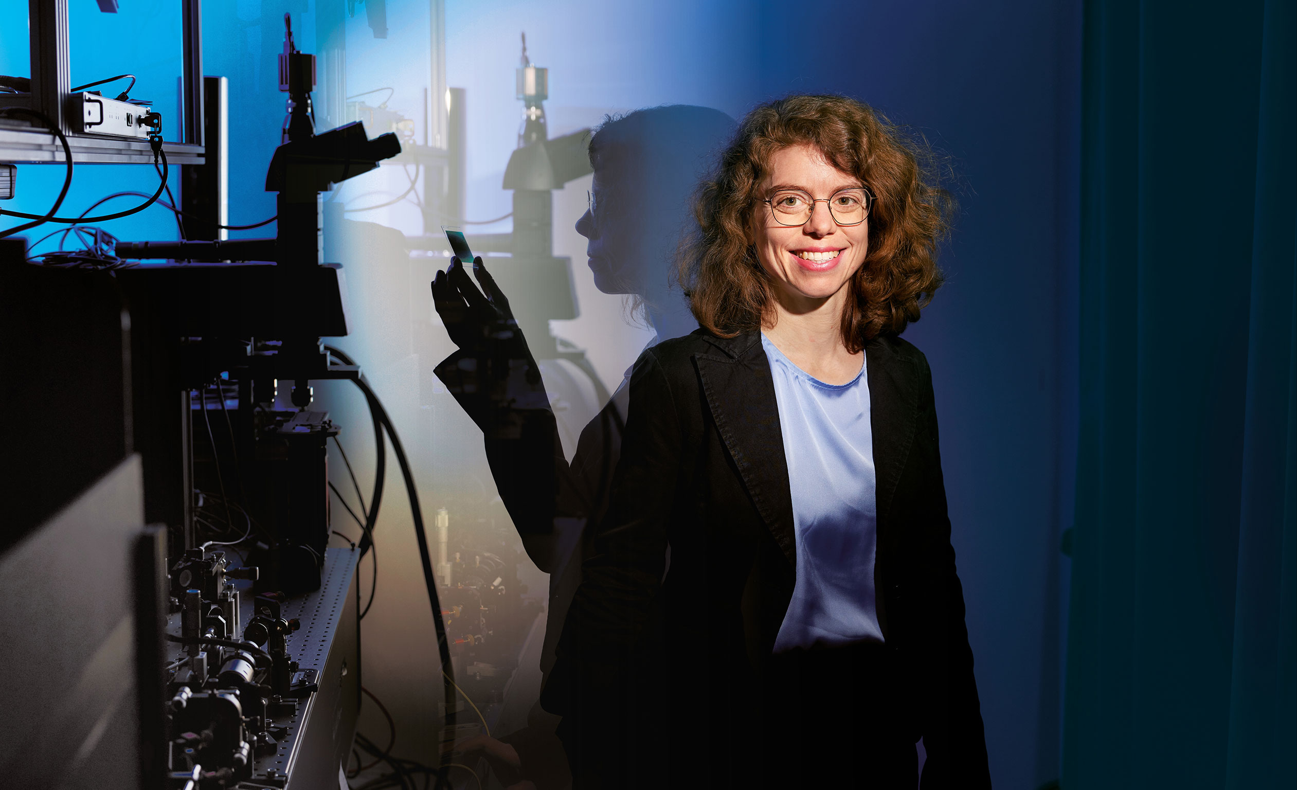
Dr. Karin Burger
© Sebastian Arlt
All tumor cells removed? Dr. Karin Burger and her team at Fraunhofer IOF are developing a quantumbased method that makes checking tissue samples after surgery faster and more efficient.

Quantum optics could offer an alternative. In the QUANCER project, which is receiving funding from the German Federal Ministry of Education and Research (BMBF), nine project partners from industry and the research sector are working together with Fraunhofer IOF on a scanning microscope that uses “undetected” light. The first step is to generate pairs of correlated light par­ticles in a photon source. These particles are complemen­tary in their quantum mechanical properties, like twins. The researchers assign them two different tasks: While one beam of light is aimed at the tissue sample, the other is captured with a camera. Because of the correlation between the particles’ frequencies, the information from the photon that has reached the sample is transferred to the other at the camera, where it is visualized – without the second photon ever coming into contact with the tissue at all. One feature in particular makes this method special: “The photons of the two light beams can even have very different wavelengths, unlocking wavelength ranges that are difficult to access,” Burger explains. “For example, one light beam in the invisible infrared range can read out specific information from the sample, such as chemical composition or changes in tissue morpholo­gy, while the other light beam lies in the visible range and can be read out by a conventional low-noise detector.” This trick could allow quantum optics to overcome the previous limits of infrared microscopy, a time-consuming and involved process.

“Our goal is a compact system for quantum imaging about the size of a shoe box, ideally compatible with a standard microscope,” Burger says. “As early as next year, the first microscope for quantum imaging in the mid-infrared range should be set up and available to examine samples using the scanning method.” Jena University Hospital plans to test the demonstrator on tumor cells at that point.